Histology
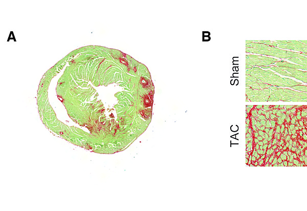
Myocardial fibrosis is assessed on a routine basis by immunohistochemistry. Paraffin-embedded or cryo-myocardial heart sections are stained for collagens (marker for fibrosis) using a Sirius Red/Fast Green staining.
In panel A, a typical 8 μm thick myocardial section is stained with Sirius Red/Fast Green. Panel B depicts representative images of control or fibrotic myocardium. The characteristic fibrosis in red is the used for quantification.